كتاب ATLAS OF HUMAN SKELETAL 2021
وصف كتاب ATLAS OF HUMAN SKELETAL 2021
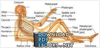
this image shows the skeleton of our body (the bones that forms and supports our body)showing:1. cranium2. atlas vertebra3. axis vertebra4. clavicle5. thoracic vertebra6. humerus7. pelvic bone8. lumbar vertebra9. cervical vertebra10. radius bone11. ulna bone12. scaphoid bone13. metacarpal bones14. phalanges of the fingers15. saddle joint of the thumb16. sacral vertebrae17. coccys vertebra18. head of femur19. femur bone20. patella21. tibia bone22. fibula bone23. metatarsal-phalangeal joint24. calcaneus boneIllustration provided by: Rolin GraphicsIn this chapter we will introduce the bones of skull and their topographic relations to each other. With the exception ofMandibula(the lower jaw), each skull- bone is connected by tight immovable joints to the bones of the neighborhood. The areas of contact between two bones are called sutures (Suturae), which represent thin junctions between the irregular interlocking edges of adjacent skull bones. In adultsSuturaeconsist of tight connective tissue. We will use many different views in this chapter to show all skull structures. To understand the views it is important to know, which position or orientation is defined as the standard. To set up a standard nomenclature for orientations of skulls in anatomy a horizontal plane was defined in Frankfort more than hundred years ago. TheFrankfort Horizontal planeis defined by tree points: the bottom margin of the left eye cavity and the top margins of the external auditory porus (see chapter 2.6. Os temporale) on both sides. Fig. 2.4.: Frankfort horizontal plane The standard anatomic perspectives are parallel or perpendicular to this (Frankfort Horizontal) plane. Perspective Explanation Norma frontalis View at the frontal outer surface of skull Norma occipitalis View at the back (posterior) outer surface of skull Norma lateralis View at the left or right outer surface of skull from side Norma verticalis View at the top outer surface of skull Norma basalis View at the inferior outer surface of skull Fig. 2.5.: Standard perspectives To describe all skull structures, it is also important to make some cuts through the skull and make the inner parts visible. In this chapter we will use the standard perspectives and additional views to show most skull structures. We will focus on major and important structures to show their topographic relations to each other. Following theSuturaeas borders and using the knowledge of this chapter’s topography it should be easy for you to learn and to describe all bones of skull. For more detailed descriptions see the following chapters which deal with separate skull bones.FHATLAS OF HUMAN SKELETAL ANATOMYJ.ARTNER ET AL. 2002, WWW.JURAJARTNER.COM 5PAGEFront view (Norma frontalis) of the skull shows the bones of the facial skull (Viscerocranium) and the frontal bone (Os frontale), which already belongs to theNeurocranium, at the top: The most prominent structures in this view are the frontal bone (Os frontale) at the top, the zygomatic arches laterally, the mandible (the lower jaw,Mandibula) at the bottom, both orbital excavations (Orbitae) below the frontal bone, the anterior nasal aperture (Apertura nasalis anterior,Apertura piriformis) located in the middle line between both orbital cavities, and the dentition, which belongs to the upper (Maxilla) and the lower jaw (Mandibula). The frontal bone(Os frontale) articulates downwards (on both sides) with the nasal bones (Ossa nasalia, sing. Os nasale), more lateral withMaxillae, the lacrimal bones (Ossa lacrimalia, sing.Os lacrimale) and with the zygomatic bones (Ossa zygomatica, sing.Os zygomaticum).Os frontaleandOssa nasaliaare joined to one another by the almost horizontal frontonasal suture (Sutura frontonasalis). The frontomaxillar suture (Sutura frontomaxillaris) unites the upper jaw (Maxilla) with the frontal bone on both sides of the frontonasal connection (suture). The lateral arched parts ofOs frontalehave contact toOs zygomaticum. Both bones form together with the upper parts ofMaxillathe exterior margins of the eye cavity (orbital cavity orOrbita) on both sides (see Fig.2.7.: Orbita). To avoid an information overload we will describe the orbital structures later in separate illustrations. At this moment it is only important to note, that the above mentioned bones extend into the orbital interior, where they form in complex connections with four other bones (Os lacrimale, Os ethmoidale, Os sphenoidale, Os palatinum) the pyramidal orbital cavities. Fig. 2.6.: Norma frontalis of skull Os frontale (1), Os nasale (2), Maxilla (3), Os zygomaticum (4), Mandibula (5), Orbita (6), Apertura nasalis ant. (7) The two nasal bones which form the bony bridge of the nose are united by the vertical internasal suture (Sutura internasalis) in the midline. The anterior nasal aperture (Apertura nasalis anteriororApertura piriformis) is located approximately in the middle of the facial cranium, below both nasal bones (Ossa nasalia) and between bothMaxillae. Divided in the middle by the nasal septum it forms the entrance into the bony nasal cavity with visible nasal shells (Conchae) and nasal septum (Septum nasi) within. The upper row of dentition (teeth) belongs to bothMaxillae, which are separated bySutura intermaxillarisunder the nasal aperture. The lower row of dentition belongs to the lower jaw (Mandibula), which is the only movable bone of skull (if the small auditory ossicles of the middle- ear are not counted).1 2 3 4 5 6 7ATLAS OF HUMAN SKELETAL ANATOMYJ.ARTNER ET AL. 2002, WWW.JURAJARTNER.COM 6PAGEFig. 2.7.: Left Orbita Os frontale (1), Maxilla (2) and Os zygomaticum (3) form the exterior margins, with Sutura frontomaxillaris (a), frontozygomatica (b) and zygo-maticomaxillaris (c) between them. Parts of Os sphenoidale (4), Os lacrimale (6), Os palatinum (7) and Os ethmoidale (5) form together with the already described bones the bottom of Orbita, separated by Sutura frontoethmoidalis (e), sphenofrontalis (d), sphenozygomatica (f), zygo-maticomaxillaris (c), ethmoido-maxillaris, frontolacrimalis, palato-maxillaris and palatoethmoidalis. Two large apertures can be seen at the bottom of the orbital cavity: Fissura orbitalis superior (Fs) connects the orbital cavity with the internal cranial cavity, passed by Nervus ophthalmicus, occulomotorius, trochlearis, Nervus abducens and Vena ophthalmica superior. Fissura orbitalis inferior (Fi), located between Os sphenoidale and the upper orbital part of Maxilla is passed by Nervus zygomaticus, Nervus infraorbitalis and their corresponding vessels .


ليست هناك تعليقات:
إرسال تعليق